1.5T MRI Scan
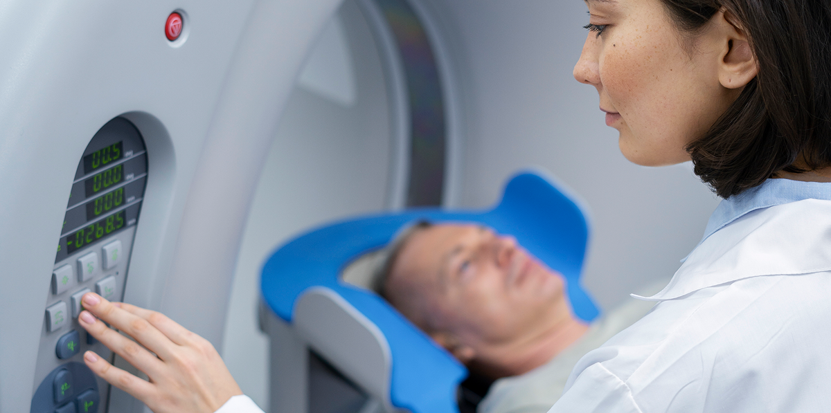
Advanced 1.5T Magnetic Resonance Imaging (MRI)
Magnetic Resonance Imaging (MRI) is a sophisticated, non-invasive diagnostic modality that uses powerful magnetic fields and radiofrequency pulses to produce high-resolution cross-sectional images of internal organs, soft tissues, bones, and neurological structures without ionizing radiation.
Comprehensive Clinical MRI Applications
Neuro-Imaging (Brain & Spine): High-definition evaluation for ischemic stroke, intracranial tumors, multiple sclerosis, aneurysms, spinal disc herniations, and nerve compression.
Musculoskeletal & Joint MRI: Detailed assessment of knee ligaments (ACL, PCL, meniscal tears), shoulder rotator cuff injuries, hip joints, and cartilage degeneration.
Abdominal & Pelvic MRI: Specialized evaluation of liver parenchyma, pancreaticobiliary system (MRCP), kidneys, and pelvic reproductive organs.
Magnetic Resonance Angiography (MRA): Non-invasive visualization of cerebral, carotid, renal, and peripheral arterial vasculature.
Patient Safety & Screening Guidelines
Because MRI utilizes strong magnetic fields, patients must undergo a thorough pre-scan safety screening. All metallic objects, watches, jewelry, hearing aids, and credit cards must be removed. Patients with cardiac pacemakers, metallic implants, or aneurysm clips must inform the radiology team prior to scheduling.
Comfort & Diagnostic Turnaround
Our imaging suites are designed for patient comfort, featuring ambient lighting, communication headsets, and wide-bore architecture. Radiologist-verified reports are accessible online shortly following scan completion.