


Understanding Immunohistochemistry (IHC)
Immunohistochemistry (IHC) is a powerful, indispensable technique in pathology, offering critical insights into the nature of diseased tissues, most notably in the diagnosis and prognosis of cancer.
The term "IHC - Each Marker" refers to the highly specific, individual application of this technique to identify and localize distinct biomarkers (antigens) within a patient's tissue sample. This process is essential for tailoring treatment plans, predicting disease behavior, and confirming diagnoses when morphology alone is insufficient.
IHC combines immunology, the study of the immune system and its antibodies, with histochemistry, the use of chemical reactions to identify substances in tissues. At its core, IHC leverages the highly specific binding property of antibodies to detect target antigens—molecular markers—that are characteristic of certain diseases or cellular processes. These antigens act as diagnostic signposts, guiding pathologists to an accurate diagnosis.
The Science Behind the Test:
The mechanism of IHC relies on a fundamental biological principle: the lock-and-key relationship between an antigen (the lock, present in the tissue) and an antibody (the key, applied in the laboratory).
Tissue Preparation: The process begins with obtaining a tissue sample, typically through a biopsy or surgical removal. This sample is carefully processed, preserved (often fixed in formalin), and embedded in a wax block (paraffin) to allow for thin sectioning. Microscopic slices of the tissue are then placed onto glass slides.
Antigen Retrieval: Because the fixation process can sometimes mask or alter the target antigens, an initial step called antigen retrieval is often necessary to expose the binding sites, ensuring the antibodies can access their targets.
Antibody Application (The "Marker"): Specific primary antibodies, designed to recognize and bind only to the target antigen of interest (the "marker"), are applied to the tissue section. The term "Each Marker" emphasizes that this test is often performed sequentially, or in parallel, for multiple different targets—for example, CD3, CD20, Ki-67, ER, PR, or HER2—depending on the diagnostic questions being asked. Each distinct antibody applied represents an individual marker analysis.
Visualization: Since antibodies themselves are not visible under a standard microscope, they are tagged with a detection system. This system usually involves a secondary antibody linked to an enzyme or a fluorescent molecule. When a chemical substrate is added, the enzyme catalyzes a reaction that produces a colored precipitate or a fluorescent signal at the exact location where the antigen-antibody complex formed. This "staining" allows the pathologist to clearly visualize the presence, location, and quantity of the target marker within the cellular context.
Clinical Utility of "IHC - Each Marker":
The utilization of IHC on a marker-by-marker basis provides essential clinical data across several key areas:
A. Diagnostic Confirmation and Subtyping of Malignancies: IHC is critical for accurately classifying tumors, especially when they are poorly differentiated (difficult to identify based on cell shape alone) or metastatic (originating elsewhere). For instance, specific markers are used to distinguish between different types of lymphomas (e.g., CD20 for B-cells, CD3/CD5 for T-cells) or to determine the primary site of an unknown metastatic tumor (e.g., TTF-1 for lung, PSA for prostate). This accurate subtyping is vital because different subtypes of cancer require vastly different treatment regimens.
B. Prognostic Information: Certain markers provide information about the likely aggressiveness or outcome (prognosis) of a disease. For example, the proliferation marker Ki-67 indicates how rapidly tumor cells are dividing, which can be an important factor in predicting disease recurrence.
C. Predictive Information (Guiding Targeted Therapy): Perhaps the most crucial modern application of IHC is determining eligibility for targeted therapies. By assessing the presence or absence of specific receptor proteins, pathologists can predict how a tumor will respond to particular drugs.
Hormone Receptors (ER/PR): In breast cancer, the presence of Estrogen Receptors (ER) and Progesterone Receptors (PR) dictates whether a patient will benefit from anti-hormonal therapy.
HER2: High expression of the HER2 protein in breast or gastric cancer suggests the patient may benefit from HER2-targeted therapies like Trastuzumab.
D. Identification of Infectious Agents and Other Non-Neoplastic Conditions: While primarily associated with cancer, IHC is also used to detect specific proteins related to infectious diseases (e.g., viral antigens) or to characterize non-cancerous conditions like autoimmune disorders.
Interpreting the Results
The analysis of IHC results is highly specialized, performed by an experienced pathologist. They examine the stained tissue under a microscope, evaluating not only whether the marker is present (positive or negative) but also:
Location: Where the stain is located within the cell (nucleus, cytoplasm, or membrane).
Intensity: The strength of the staining reaction.
Pattern: The distribution of the positive cells within the tissue.
Quantification: For some markers (like ER, PR, and Ki-67), the percentage of positive cells is counted and reported, often leading to a scoring system (e.g., Allred score or H-score). The interpretation of "IHC - Each Marker" is always integrated with other clinical data and traditional microscopic examination (H&E staining) to arrive at a comprehensive and precise diagnosis.
Summary:
The "IHC - Each Marker" analysis is an indispensable tool in modern precision medicine. It transforms passive tissue observation into an active molecular investigation. By individually targeting and visualizing specific antigens, this test provides the molecular fingerprints of disease, enabling physicians to make accurate diagnoses, determine the aggressiveness of a condition, and select the most effective, personalized therapeutic strategies for improved patient outcomes.
Frequently Asked Questions (FAQs)
Is a blood sample required for the IHC test?
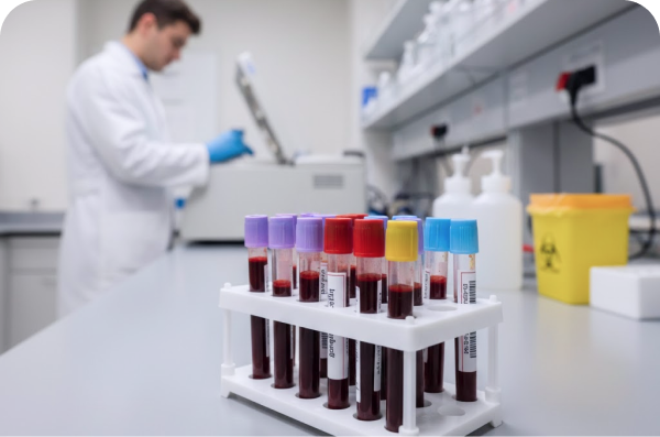
No, the IHC – Single Marker test is performed on a tissue sample, not a blood sample. The tissue is obtained through a biopsy or surgical procedure prior to IHC analysis.
How long do results take?
IHC usually adds about one extra day to routine tissue processing, with total turnaround typically ranging from 2 to 10 days depending on the complexity of the marker and whether the sample is being processed in-house or referred to a specialised reference laboratory.
What is the difference between IHC – Single Marker and an IHC Panel?
A single marker test applies one targeted antibody to answer one specific clinical question such as confirming ER status. An IHC panel applies multiple antibodies simultaneously, typically used when the tissue of origin is unknown or when a broad differential diagnosis needs to be resolved. The single marker format is chosen when the diagnostic question is already well-defined and one confirmatory result is all that is required.
Is IHC the same as a genetic test?
No, IHC detects the presence and quantity of a protein within tissue cells, while genetic tests such as FISH, PCR, or next-generation sequencing analyse DNA or RNA.
Can IHC be performed on old tissue blocks?
Yes, IHC can generally be performed on archival FFPE (formalin-fixed, paraffin-embedded) tissue blocks stored for several years, provided the original fixation was adequate. The laboratory will retrieve and section the stored block for analysis.
How accurate is the IHC – Single Marker test?
IHC is highly accurate, often above 90–95% for well-established markers, when performed using quality-controlled reagents and interpreted by experienced pathologists at accredited diagnostic facilities.
Does a positive IHC result always confirm cancer?
Not necessarily IHC positivity for a given marker indicates the presence of that specific protein in the tissue. Whether this confirms malignancy depends entirely on which marker was tested and the clinical context.