

HRCT Chest in Secunderabad
Also called: Chest CT, Lung scan, CT Thorax
- SAMPLE
- Not required
- Report
- Same or next day
- PREPARATIONS
- Fasting 4–6 hrs
The Scan at a glance
- What it checks
- SURGICAL PROFILE WITH CT CHEST test is a clinically significant diagnostic investigation used to evaluate, screen, or monitor underlying health conditions. It provides actionable insights that support early diagnosis, treatment planning, and long-term disease management.
- Report delivery
- For report delivery timelines, please contact the front desk or call +91 93901 10089.
- Test for
- Men, Women
- Age group
- 20
- Commonly advised for
- Persistent cough, shortness of breath, chest pain
- Home collection
- No
What is a Complete Blood Count?
Know More About This Test
The Surgical Profile with CT Chest is a comprehensive pre-operative evaluation combining blood investigations and imaging. It ensures that a patient is medically fit for surgery and identifies any hidden risks such as lung infections, cardiac strain, coagulation issues, or metabolic abnormalities. The CT chest component provides detailed imaging of lungs and mediastinum, helping surgeons plan anesthesia and surgical approach safely.
Why Is This Profile Recommended?
You may need this profile to:
- Conduct a thorough pre-operative safety assessment that evaluates blood health, organ function, coagulation, infectious disease status, and detailed lung anatomy in a single, coordinated investigation
- Assess the function of the lungs, detect abnormal growth in the lungs, and detect obstruction to airflow through the lungs before a patient is exposed to general anaesthesia, which places significant demands on respiratory reserve
- Identify any underlying pulmonary pathology — such as interstitial lung disease, emphysema, pulmonary fibrosis, or endobronchial lesions — that could complicate intraoperative ventilation or post-operative recovery
- Screen for transmissible infections (HIV, HBsAg, HCV) to protect both the patient and the surgical team
- Detect uncontrolled blood sugar, electrolyte imbalances, coagulation abnormalities, or anaemia that require correction before the procedure date
- Evaluate cardiac rhythm via ECG before administering anaesthesia, and assess renal and hepatic function to ensure the body can safely process anaesthetic agents and surgical medications
Symptoms or Conditions That May Require This Profile
This profile is particularly indicated for patients who:
- Are scheduled for a major elective or emergency surgical procedure and have a history of chronic respiratory disease such as COPD, asthma, pulmonary fibrosis, or recurrent chest infections
- Have a significant smoking history, as this substantially increases the risk of occult pulmonary pathology that may not be apparent on a routine chest X-ray
- Present with symptoms such as persistent cough, breathlessness on exertion, haemoptysis, or unexplained weight loss that require detailed pulmonary evaluation before surgery
- Have occupational exposure to dust, asbestos, silica, or other inhaled toxins associated with occupational lung disease
- Are above the age of 50 and are undergoing thoracic, cardiac, or upper abdominal surgery, where pulmonary reserve directly affects surgical and anaesthetic risk
- Have been treated for tuberculosis and require assessment of residual lung damage prior to surgery
- Have autoimmune conditions such as rheumatoid arthritis, scleroderma, or lupus, which are known to cause interstitial lung disease that requires characterisation before anaesthesia
How Is the Test Performed and Prepared For?
Blood samples are collected for laboratory analysis and CT chest imaging is performed. Fasting may be required for blood tests. Inform about contrast allergy if contrast is used. Remove metal objects before CT.
What Does the Surgical Profile With HRCT Chest-II Assess?
Blood and Laboratory Investigations:
- Complete Blood Picture (CBP): Haemoglobin, RBC, WBC, platelets, and differential count screening for anaemia, infection, and clotting disorders
- Blood Grouping and Rh Typing: Identifies ABO blood group and Rh factor for emergency transfusion preparedness
- Prothrombin Time with INR: Assesses coagulation and surgical bleeding risk
- Liver Function Tests (LFT): Evaluates hepatic health and the liver's capacity to process anaesthetic agents and surgical medications
- Serum Creatinine: Assesses kidney function and ability to clear anaesthetic drugs and contrast agents
- Random Blood Sugar (RBS): Screens for uncontrolled diabetes before surgery
- Serum Potassium: Detects electrolyte imbalances that can affect cardiac rhythm during anaesthesia
- HIV 1 & 2, HBsAg, Anti-HCV: Infectious disease screening for patient and surgical team safety
- Resting ECG: Evaluates cardiac rhythm and detects arrhythmias or ischaemic changes before anaesthesia
HRCT Chest:
- A high-resolution imaging procedure that uses narrow beams of X-rays to create detailed cross-sectional images of the lung anatomy, assessing lung function, detecting abnormal growths, and identifying obstruction to airflow through the lungs
- Evaluates the lung parenchyma for interstitial disease, emphysema, ground-glass opacities, consolidation, nodules, and fibrosis
- Assesses the airways, pleural spaces, mediastinum, and hilar structures
- Identifies active or healed pulmonary infections including tuberculosis and fungal disease
- Detects early interstitial lung abnormalities that may represent early-stage fibrosing lung disease requiring characterisation before surgery and anaesthesia
What Do the Results Mean?
Blood results:
- Normal parameters across all blood investigations indicate that the patient's haematological, metabolic, coagulation, and infectious disease status is compatible with safe surgical and anaesthetic management
- Abnormal values in specific parameters will indicate which organ system requires correction or further specialist evaluation before surgery proceeds
HRCT Chest:
- Normal HRCT: Lung parenchyma, airways, pleura, and mediastinum appear within normal limits no significant pulmonary pathology identified. Surgery may proceed from a respiratory standpoint
- Ground-glass opacities: Areas of mildly increased lung density that may represent early infection, inflammation, or interstitial disease. Clinical correlation and pulmonologist review are required
- Consolidation: Dense filling of air spaces suggesting active infection, organising pneumonia, or malignancy requires further evaluation before elective surgery
- Nodules: Discrete rounded densities that may be benign or malignant characterisation and follow-up planning are essential, particularly if the nodule is new or growing
- Reticular opacities and honeycombing in a peripheral, basal distribution suggest Usual Interstitial Pneumonia (UIP) or idiopathic pulmonary fibrosis conditions with significant implications for post-operative respiratory management
- Tree-in-bud pattern: Represents dilated, mucus-filled centrilobular bronchioles and almost invariably indicates endobronchial spread of infection such as tuberculosis, Mycobacterium avium complex, or bacterial bronchopneumonia requires infectious disease evaluation before surgery
Lifestyle Tips to Support Safe Surgical Preparation
- Follow all fasting and medication instructions provided by the surgical team precisely.
- Quit smoking or significantly reduce smoking before the procedure date smoking impairs wound healing.
- Control blood sugar carefully in the days leading up to surgery.
- Avoid alcohol for at least 48 hours before the procedure, as alcohol affects coagulation, liver function, and the metabolism of anaesthetic agents
- Maintain prescribed medications for chronic conditions such as hypertension, asthma, or thyroid disease.
- Attend all pre-operative appointments, investigations, and specialist consultations without delay.
Frequently Asked Questions (FAQs)
- Is CT chest mandatory before surgery? Depends on surgery type; required for major thoracic or oncologic procedures.
- Does CT involve radiation? Yes, but controlled; low-dose radiation used for diagnostic imaging.
- Can surgery proceed if abnormal? Sometimes after correction; treatable issues can be addressed first.
- Is contrast always used? Not always; depends on what needs evaluation.
- How long does the process take? Same day usually; results available within 24-48 hours.
Who should take this test
HRCT of the chest is a high-resolution scan that shows fine detail in the lung tissue. It is advised if:
- You have a long-standing cough or breathlessness
- Your doctor suspects a lung condition affecting the small airways
- An earlier X-ray or CT needs finer detail
- You have a known lung disease under follow-up
- You are being assessed before a chest procedure
HRCT Chest in Secunderabad
Worried about your HRCT Chest scan? Tapadia Diagnostic Centre in Secunderabad offers AERB-licensed HRCT Chest. Chest CT, Lung scan, CT Thorax is commonly prescribed for Persistent cough, shortness of breath, chest pain and suitable for 20 Men, Women. Pay at the centre with no advance payment.
Frequently asked questions
Related Scans in Secunderabad




Centers You Can Choose To Visit
Vijayawada
- H.no 29-27-22, Suryanarayana street, suryaraopet vijayawada-520002
- Services: Lab
- Branch Incharge: Mr. Akhil : +91 6300387321
Get Direction
KPHB COLONY
- MIG H1, Plot No 273, above Visakhapatnam Coop Bank, Kukatpally Housing Board Colony, K P H B Phase 1, Kukatpally, Hyderabad, Telangana 500072
- Richards-9866362080
- Services - Lab, Digital X-Ray. ECG
Get Direction
Tapadia Diagnostic Center Bengaluru
- Sahakarnagar Main Rd, G Block, Sahakar Nagar, Byatarayanapura, Bengaluru, Karnataka 560092
- All facilities Available
- Branch Incharge: Mohammad Sarfaraz :+91 79896 92437
Get Direction
TAPADIA DIAGNOSTIC CENTRE (Main Branch)
- Main Branch
- 1-7-1072/A, RTC X Roads, Opp Saptagiri Theatre, Hyderabad, Telangana, India 500020.
- All facilities Available
- Give call at -8305555999
- Alternate Number -9390110098
Get Direction
SECUNDERABAD
- St. Thomas Spg Church Telugu Pastorate, near Sitara Hotel, Complex, Palika Bazar, Monda Market, Shivaji Nagar, Secunderabad, Telangana 500003
- Services: Lab, ECG
- Ravi-9100577378
Get Direction
Chhatrapati Sambhajinagar (Aurangabad)
- Bansilal Nagar, Station Road, Chhatrapati Sambhajinagar (Aurangabad).
- Services: MRI, CT Scan, Ultrasound, Lab, X Ray, ECG
- Amar Mundada: 94055 85858
Get Direction
HRCT Chest in Secunderabad
Rated 4.6 on Google From 6,477 patient reviews across our Hyderabad centres.
Every report, kept for you Past reports stay in your account and can be downloaded again.
NABL accredited Every result reviewed by a qualified pathologist before it reaches you.
Four decades of practice A family-run diagnostic centre, trusted by doctors and households since 1989
Medically Reviewed by Qualified Biochemists

HRCT Chest in Other Cities
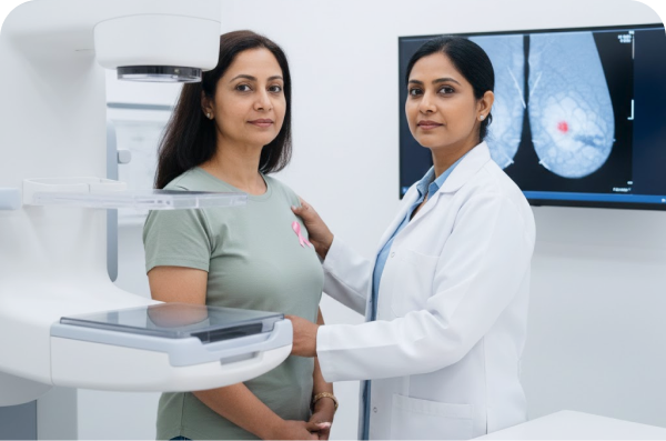
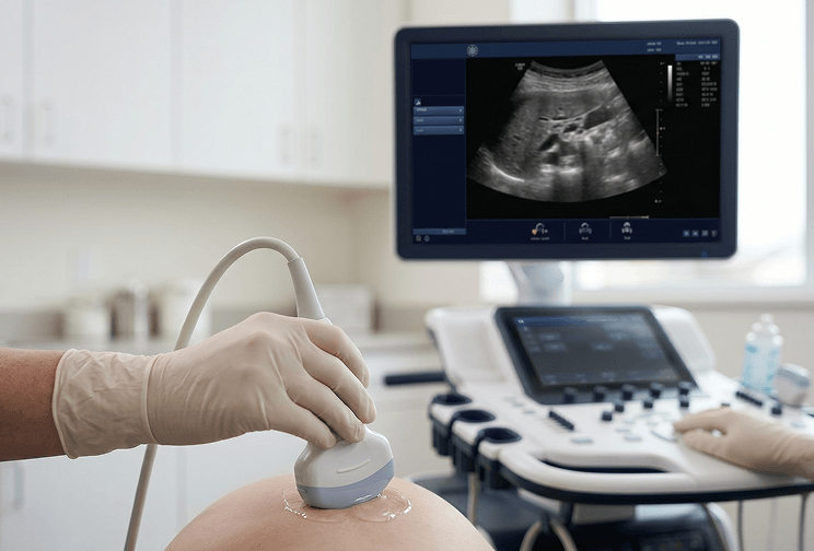
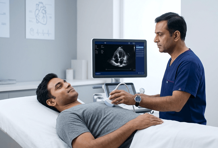
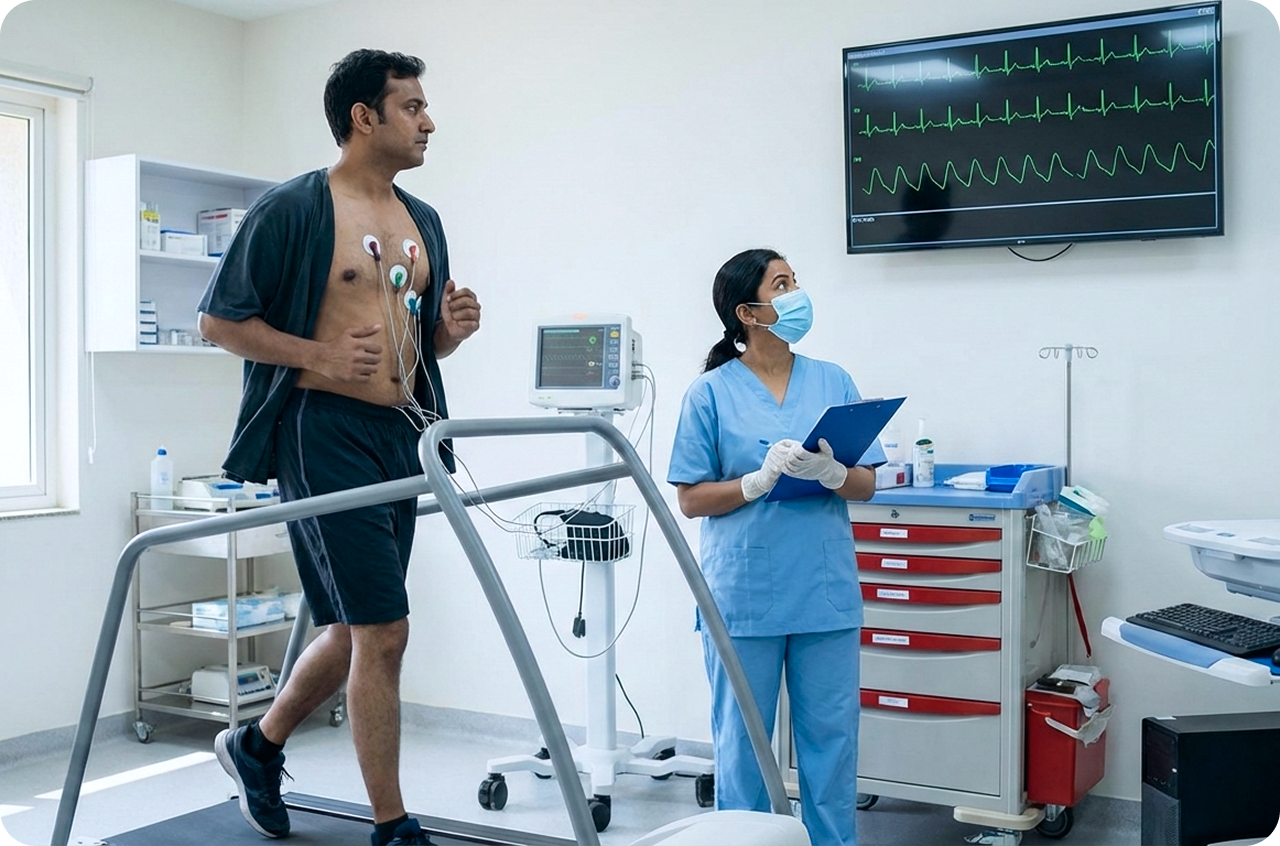
MRI Knee - Both Joints in SecunderabadMRI Knee - Left Joint in SecunderabadMRI Knee - Right Joint in SecunderabadMRI C Spine in SecunderabadMRI LS Spine in SecunderabadMRI Brain in SecunderabadPulmonary Function Test in SecunderabadMRI - Brain/Spine/Knee in SecunderabadElectrocardiogram (ECG) in SecunderabadMammogram in SecunderabadChest X-ray in SecunderabadAbdominal Ultrasound in Secunderabad2D Echo in SecunderabadExercise Stress Test in SecunderabadPrenatal Ultrasound in SecunderabadX-Ray KUB in SecunderabadChest CT Scan in SecunderabadEEG in SecunderabadCT Head in SecunderabadHRCT Chest in SecunderabadAnomaly Scan in Secunderabad

 Made with Levo