
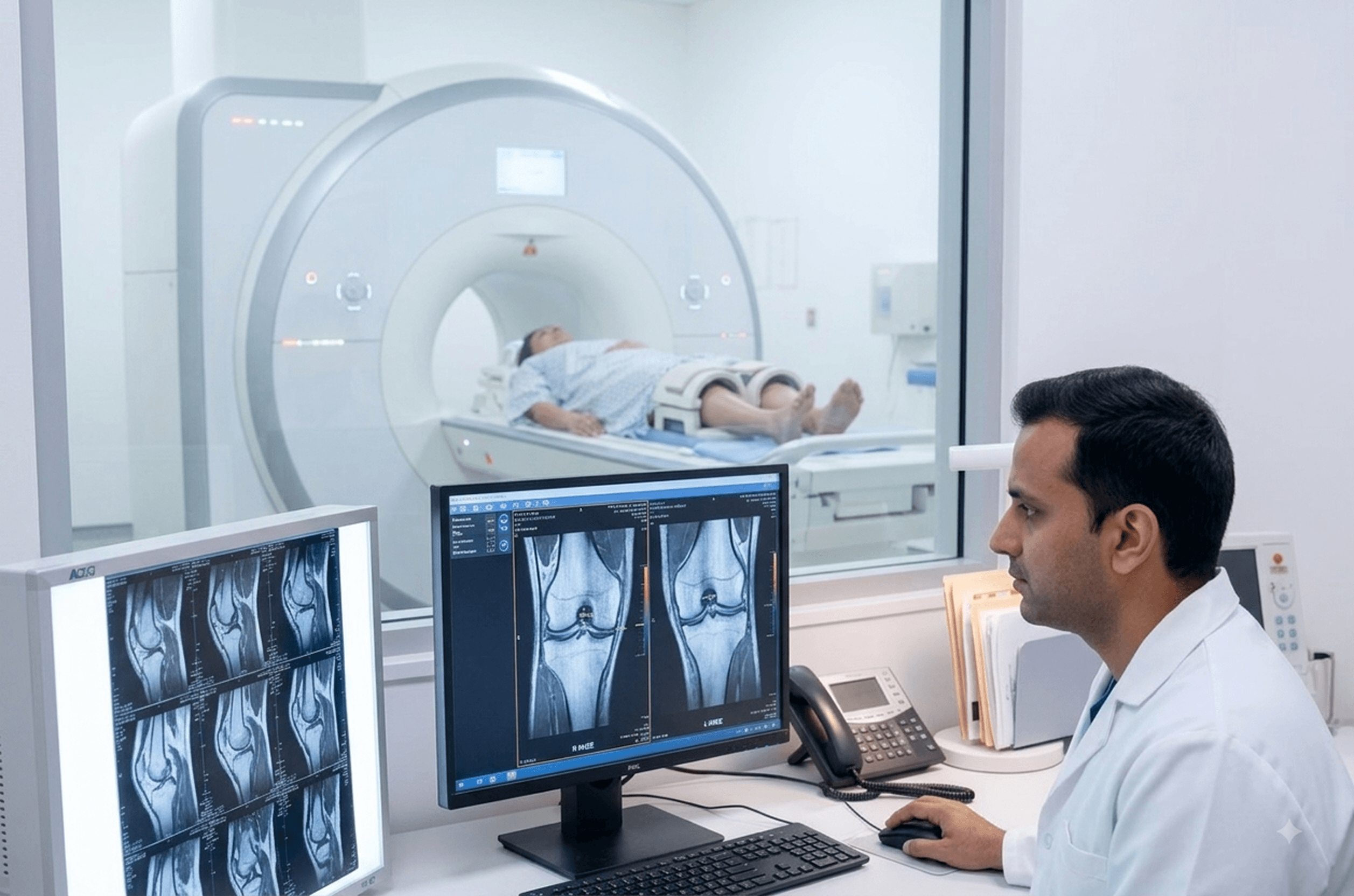

MRI - Brain/Spine/Knee in Warangal
Also called: Nuclear magnetic resonance imaging, NMR imaging, Scan
- SAMPLE
- Not required
- Report
- Same or next day
- PREPARATIONS
- Remove metal and jewellery
The Scan at a glance
- What it checks
- An MRI of the brain, spine, or knee is a non-invasive diagnostic imaging test that uses powerful magnets and radio waves to create detailed images of these areas to help diagnose injuries, tumors, cysts, or other abnormalities.
- Report delivery
- For report delivery timelines, please contact the front desk or call +91 93901 10089.
- Test for
- Men, Women
- Age group
- All
- Commonly advised for
- Knee pain, swelling, locking, instability, stiffness
- Home collection
- No
What is a Complete Blood Count?
Understanding Magnetic Resonance Imaging (MRI)
Magnetic Resonance Imaging (MRI) is a cutting-edge, non-invasive imaging technology that uses a powerful magnetic field, radio waves, and a computer to produce detailed pictures of organs, soft tissues, bone, and virtually all other internal body structures. Unlike X-rays or CT scans, an MRI does not use ionizing radiation, making it a safe diagnostic tool for many conditions.
The procedure is particularly valuable for examining the body's major joints and the central nervous system, which includes the brain and spine. The high-resolution images generated by an MRI allow physicians to assess injuries, diagnose diseases, monitor treatment effectiveness, and plan procedures with great precision.
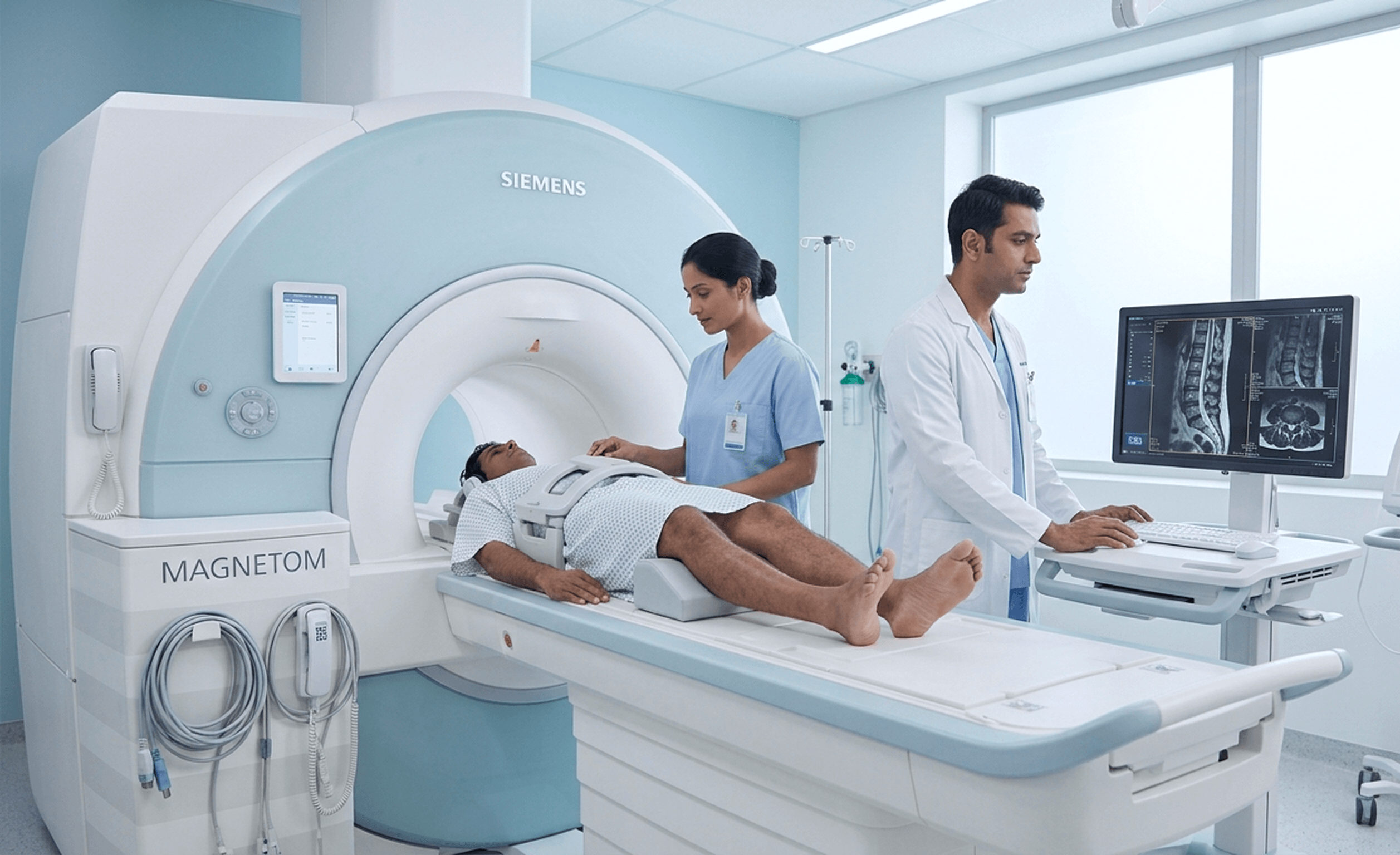
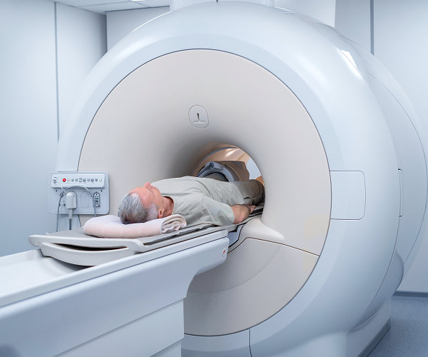
What is an MRI? MRI technology relies on the natural magnetic properties of water molecules within the body. When a patient is placed inside the MRI scanner (a large, tunnel-shaped machine), the strong magnetic field temporarily aligns these water molecules. Radio waves are then pulsed through the body, knocking the aligned molecules out of position. As the molecules snap back into alignment, they emit signals that are detected by the scanner. A computer processes these signals to create cross-sectional images, or "slices," of the targeted area.
In some cases, a contrast agent, often a substance called gadolinium, is injected intravenously. This substance enhances the clarity of the images, especially for blood vessels, tumors, and inflammation.
Preparing for Your MRI: Preparation is critical to ensure both the safety of the patient and the quality of the images. Because the MRI machine uses an extremely strong magnet, no magnetic metal objects must enter the scanner room.
Before the exam, you will be asked to complete a detailed safety screening form. You will typically be required to change into a hospital gown and remove all metal objects, including:
- Jewelry, watches, and hairpins
- Eyeglasses and hearing aids
- Wigs and dentures
- Clothing with metal snaps, zippers, or underwire
Certain implanted medical devices containing metal may prevent you from having an MRI. It is vital to inform your physician and the MRI technologist if you have any of the following:
- Pacemakers or implantable cardioverter-defibrillators (ICDs)
- Intracranial aneurysm clips
- Cochlear implants
- Metallic joint prostheses (depending on type and certification)
- Metal fragments from accidents or surgery
You should also discuss any history of kidney problems, asthma, or allergies, particularly if contrast dye is required for your exam.
Specific Applications of MRI:
MRI of the Brain and Spine An MRI of the brain and spine provides unparalleled detail of the central nervous system. It is often used to diagnose and evaluate:
- Neurological disorders: Multiple sclerosis (MS), stroke, dementia, and epilepsy.
- Structural abnormalities: Congenital malformations and hydrocephalus.
- Tumors and cysts: Identifying their size, location, and relationship to surrounding tissues.
- Spinal issues: Herniated discs, spinal cord compression, nerve root impingement, and infection or inflammation along the spine.
During a brain or spine MRI, the patient lies on a movable table that slides into the scanner. The exam is painless but requires the patient to remain perfectly still to avoid blurring the images. The procedure typically takes 30 to 60 minutes.
MRI of the Knee: The knee joint is complex, involving bone, cartilage, ligaments, tendons, and muscles. In combination with conventional X-rays, MRI is the preferred method for examining the knee because it provides clear images of soft tissues that X-rays cannot capture.
A knee MRI is performed to diagnose or evaluate:
- Injuries: Tears in ligaments (like the ACL or PCL), tendons, or the meniscus (cartilage).
- Inflammation and swelling: Detecting conditions like arthritis or bursitis.
- Internal bleeding or fluid accumulation within the joint.
- Bone issues: Identifying fractures, infections, or tumors.
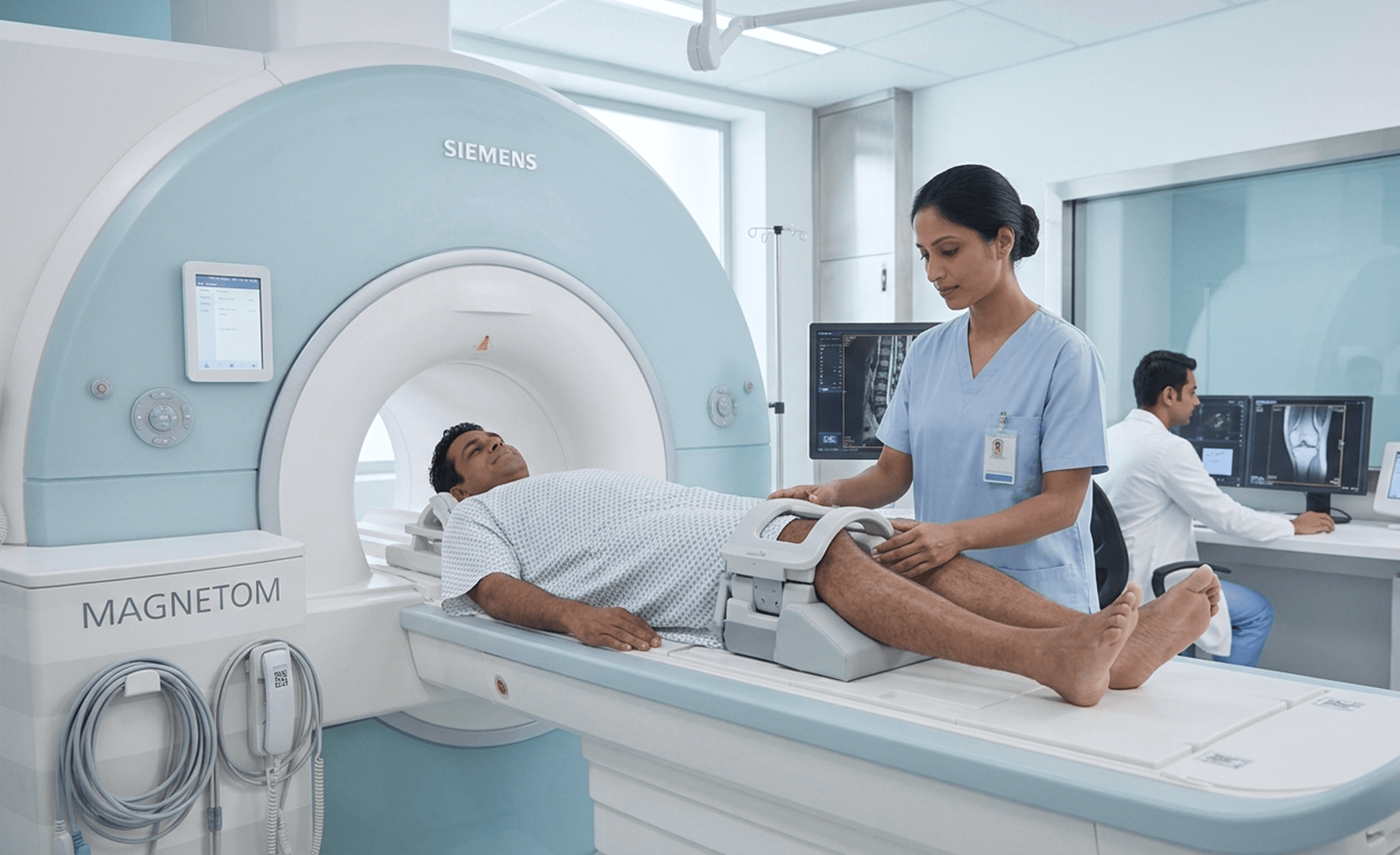
The patient is positioned comfortably on the table, and a specialized coil may be placed around the knee to enhance image quality.
What to Expect During the Exam: Once inside the scanner, you will hear loud tapping or thumping noises as the machine generates the radio frequency pulses. Hearing protection, such as earplugs or headphones, will be provided. The technologist will monitor you from an adjacent room and can communicate with you through an intercom system. You may also be given a call button to press if you feel uncomfortable.
It is important to remember that MRI is a safe, radiation-free procedure. Although the scanner environment can be confining, the detailed information it provides is essential for accurate diagnosis and effective treatment planning.
Cost and Insurance: MRI costs vary widely based on geographic location, the specific area of the body being scanned, and the facility (hospital versus outpatient center). Without insurance, the cost can range significantly. Patients with insurance should consult their provider to understand their specific coverage, including deductibles and copays, as insurance coverage can significantly reduce out-of-pocket expenses.
Who should take this test
This MRI covers the brain, spine or knee as advised by your doctor, using magnets rather than radiation. It is advised if:
- You have persistent headaches, dizziness or seizures
- You have back or neck pain travelling into a limb
- You have knee pain, swelling or locking after an injury
- You have numbness, tingling or weakness
- An earlier X-ray did not explain your symptoms
MRI - Brain/Spine/Knee in Warangal
Worried about your MRI - Brain/Spine/Knee scan? Tapadia Diagnostic Centre in Warangal offers AERB-licensed MRI - Brain/Spine/Knee. Nuclear magnetic resonance imaging, NMR imaging, Scan is commonly prescribed for Knee pain, swelling, locking, instability, stiffness and suitable for All Men, Women. Pay at the centre with no advance payment.
Frequently asked questions
Related Scans in Warangal



Centers You Can Choose To Visit
Vijayawada
- H.no 29-27-22, Suryanarayana street, suryaraopet vijayawada-520002
- Services: Lab
- Branch Incharge: Mr. Akhil : +91 6300387321
Get Direction
KPHB COLONY
- MIG H1, Plot No 273, above Visakhapatnam Coop Bank, Kukatpally Housing Board Colony, K P H B Phase 1, Kukatpally, Hyderabad, Telangana 500072
- Richards-9866362080
- Services - Lab, Digital X-Ray. ECG
Get Direction
Tapadia Diagnostic Center Bengaluru
- Sahakarnagar Main Rd, G Block, Sahakar Nagar, Byatarayanapura, Bengaluru, Karnataka 560092
- All facilities Available
- Branch Incharge: Mohammad Sarfaraz :+91 79896 92437
Get Direction
TAPADIA DIAGNOSTIC CENTRE (Main Branch)
- Main Branch
- 1-7-1072/A, RTC X Roads, Opp Saptagiri Theatre, Hyderabad, Telangana, India 500020.
- All facilities Available
- Give call at -8305555999
- Alternate Number -9390110098
Get Direction
SECUNDERABAD
- St. Thomas Spg Church Telugu Pastorate, near Sitara Hotel, Complex, Palika Bazar, Monda Market, Shivaji Nagar, Secunderabad, Telangana 500003
- Services: Lab, ECG
- Ravi-9100577378
Get Direction
Chhatrapati Sambhajinagar (Aurangabad)
- Bansilal Nagar, Station Road, Chhatrapati Sambhajinagar (Aurangabad).
- Services: MRI, CT Scan, Ultrasound, Lab, X Ray, ECG
- Amar Mundada: 94055 85858
Get Direction
MRI - Brain/Spine/Knee in Warangal
Rated 4.6 on Google From 6,477 patient reviews across our Hyderabad centres.
Every report, kept for you Past reports stay in your account and can be downloaded again.
NABL accredited Every result reviewed by a qualified pathologist before it reaches you.
Four decades of practice A family-run diagnostic centre, trusted by doctors and households since 1989
Medically Reviewed by Qualified Biochemists

MRI - Brain/Spine/Knee in Other Cities
MRI Knee - Both Joints in WarangalMRI Knee - Left Joint in WarangalMRI Knee - Right Joint in WarangalMRI C Spine in WarangalMRI LS Spine in WarangalMRI Brain in WarangalPulmonary Function Test in WarangalMRI - Brain/Spine/Knee in WarangalElectrocardiogram (ECG) in WarangalMammogram in WarangalChest X-ray in WarangalAbdominal Ultrasound in Warangal2D Echo in WarangalExercise Stress Test in WarangalPrenatal Ultrasound in WarangalX-Ray KUB in WarangalChest CT Scan in WarangalEEG in WarangalCT Head in WarangalHRCT Chest in WarangalAnomaly Scan in Warangal

 Made with Levo